
|
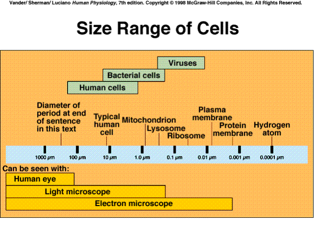
Cell Size
|
|
|
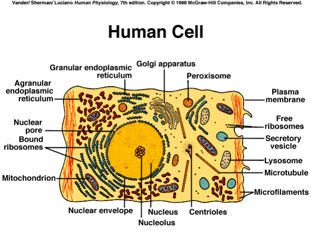
Human Cell
|
|
A. Nucleus
– contains the chromosomes; often in center
B. Cytoplasm
|
Cytoplasm and Cytosol
|

|
|
Cytoskeletal Filaments
|

|

Cell organelles are sub-units of the cell that cannot function if removed from the cell; perform specific functions
|
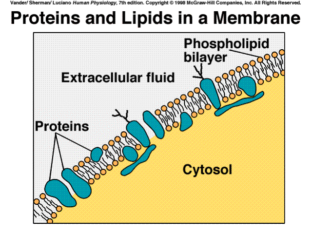
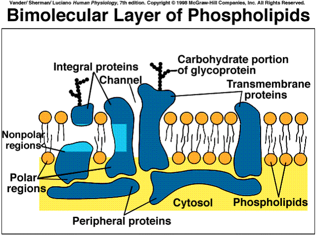
Membrane
|
|
It is the outer boundary of the cell and is responsible for regulating whether substances enter or leave the cell; semi-permeable; barrier for material to move into and out of the cell
|
Phospholipid
|
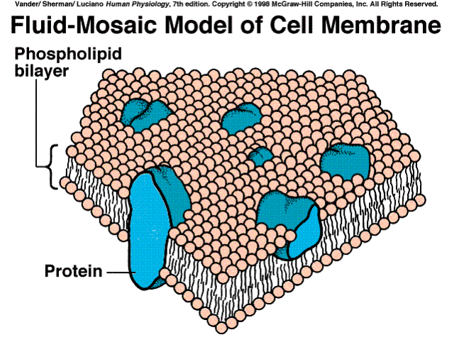
Fluid Mosaic
|
|
|
|
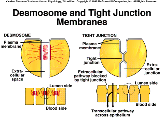
Desmosome
|
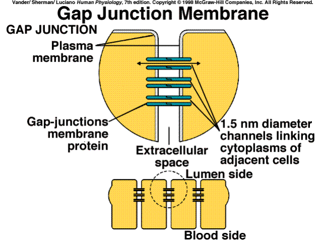
Gap Junction
|
|
|
|
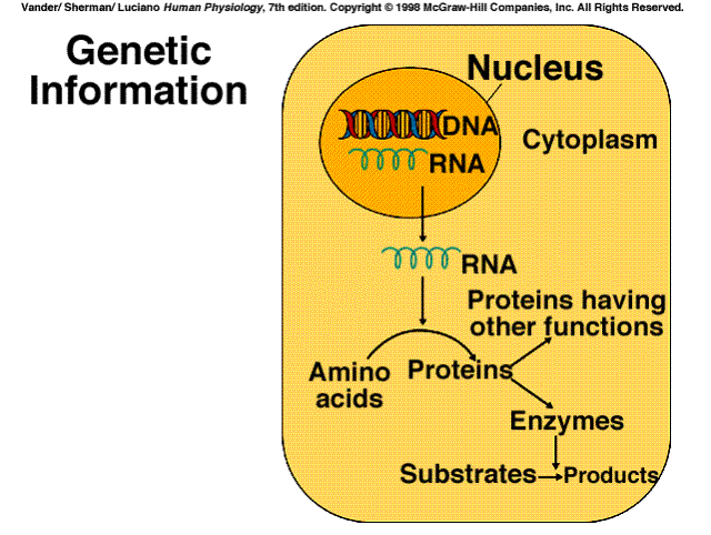
Genetic Information
|
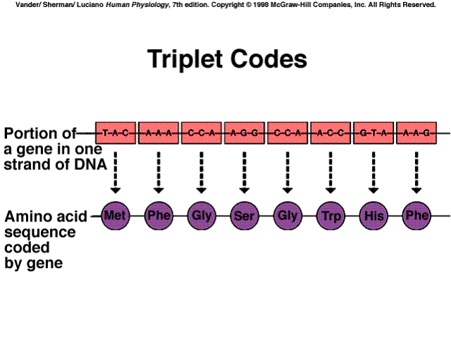
Genetic Code
|
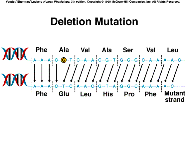
Deletion Mutation
|
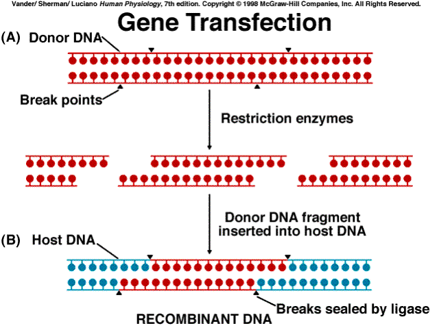
Transfection
|
|
|
|
|
|
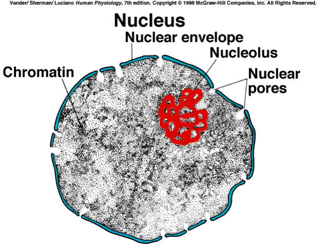
Nucleolus
|
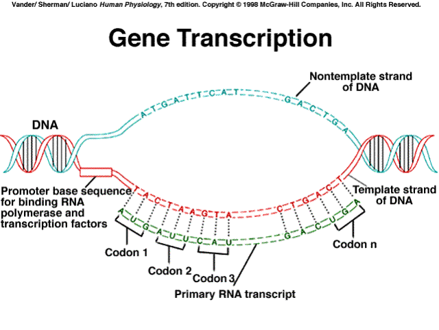
Gene Transcription
|
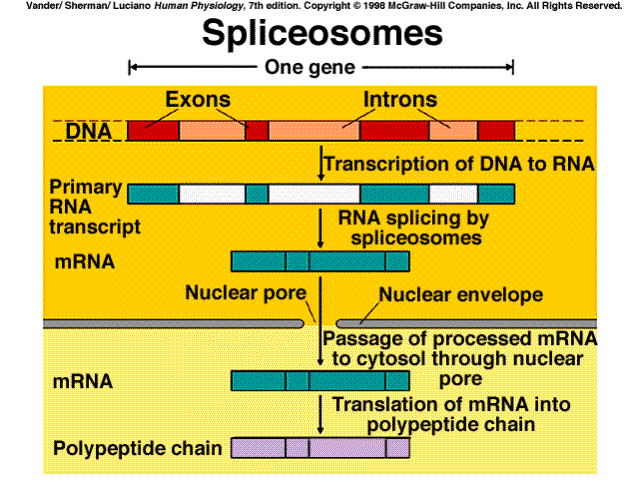
Splicesomes
|
|
|
|
|
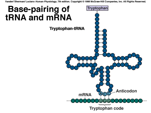
Base Pairing
|
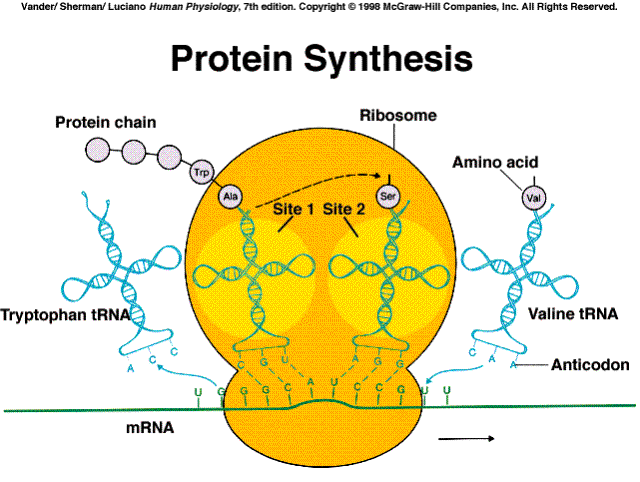
Protein Synthesis
|
Ribosomes
|
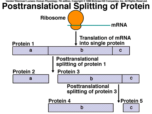
Protein Splitting
|
|
|
|
|
|
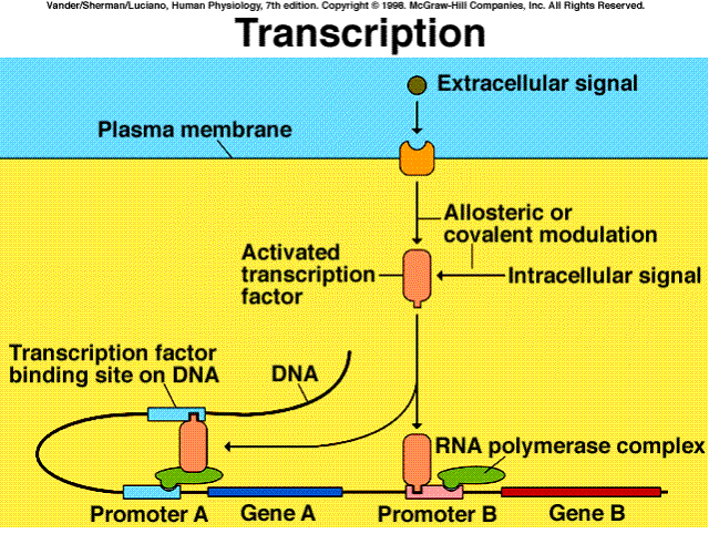
Transcription
|
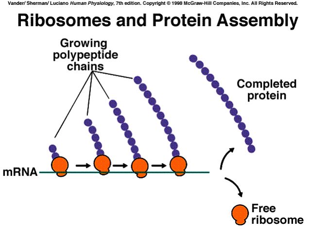
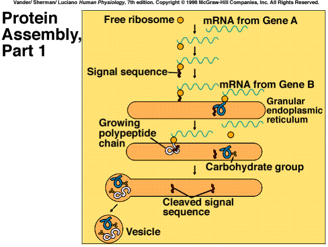
Protein Assembly 1
|
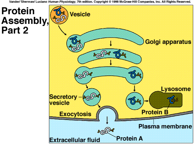
Protein Assembly 2
|
|
|
|
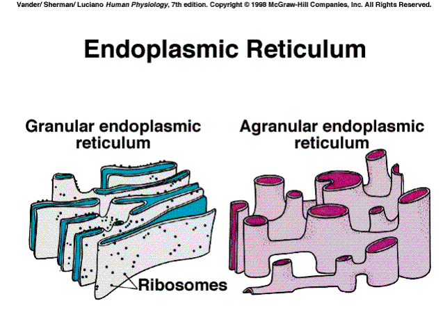
– platforms that provide a surface location for the assembly of amino acids into proteins. Ribosomes are constructed of protein and ribosomal ribonucleic acid (r-RNA) and are attached to endoplasmic reticulum [an internal cell membrane]. When ribosomes are attached to endoplasmic reticulum, the endoplasmic reticulum is called rough or granular endoplasmic reticulum. Also present are proteins that serve as enzymes that allow the linkage of amino acids to form a polymer of amino acids [protein]
|
Endoplasmic Reticulum
|
|
|
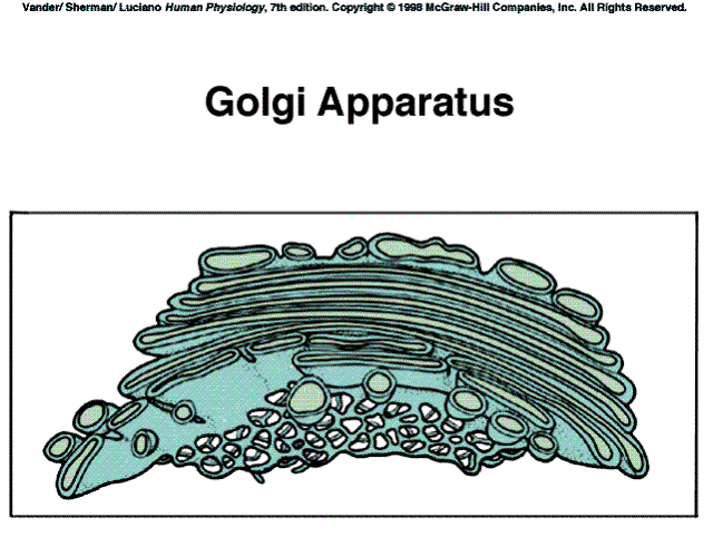
Golgi Apparatus
|
|
|
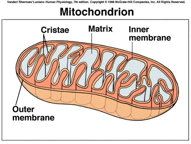
Mitochondria
|
|
Mitochondria are rod-shaped organelles with a double outer membrane. The mitochondria contain a special set of enzymes that enable the creation of large amounts of an energy storage molecule called adenosine triphosphate (ATP). Oxygen is required to be present for the enzyme systems to function in the mitochondria.
Lysosomes are small vesicles containing acid and digestive enzymes that breakdown bacteria, debris from dead cells, and non-functioning organelles from the same cell.
Peroxisomes are similar to lysosomes except they contain enzymes.These enzymes are primarily hydrogen peroxide, which is toxic to the cell. Within the cell, there is a variety of filaments including those necessary for cell division and those necessary for movement of the cell.
Mitosis is the process which produces cell "clones". It is multiplication through division; one cell divides into two identical clone cells and has increased the total cell population. Using the poweres of multiplication; 2, 4, 8, 16, 32, 64, 128, 256, 512, 1024, 2048, ... it does not take long to produce millions and billions of cells. We call the uncontrolled proliferation of cells in this manner "cancer". Normally, we have a myriad of controls which limit and specify cell growth and division.
|
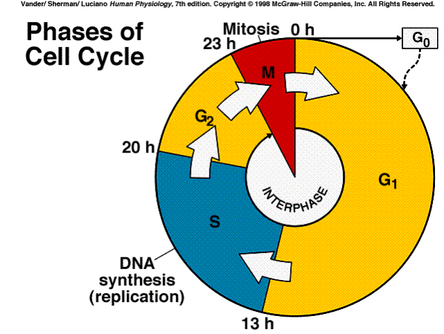
Cell Cycle
|
|
The "Cell Cycle" is the set of events that a cell typically transverses during its life. After a cell separates from it sibling daughter cell it enters a stage known as G1. (Which is the first growth or gap period.) Some cells tend to hang at this point forever, e.g., liver, nerve, and muscle cells. However, given proper stimuli, they will move on to the next stage.
The second stage is the "S" stage where the synthsis of DNA occurs. Once a cell passes from G1 to S stage, it will divide.
|
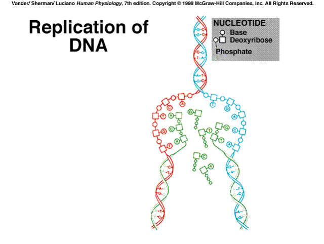
Replication
|
|
Subsequently the cell proceeds through a second "Growth" stage where it performs the rest of the tasks required in preparation for the actual division, Mitosis.
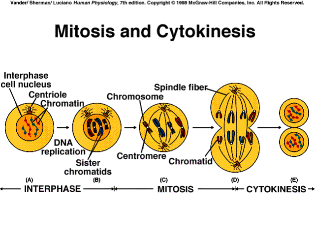
Mitosis can generally be divided into four phases, Prophase, Metaphase, Anaphase, and Telophase.
During prophase, the chromosomes condense and the nuclear membrane dissappears.
During metaphase, the chromsomes align along a center axis of the cell and the astrocytes align on either side of the equator.
During anaphase, the chromosomes are pulled apart and one set is segregated to each half of the cell.
During telephase, the cell is pinched into two around the equator, the nuclear membrane reforms, the chromosomes disperse, and we are left with two daughter cells.
|
Mitosis
|
|
The majority of cells are 10 to 20 mm, but the range of cell size is from 2 mm to 120 mm. However, a nerve cell may be a meter in length. It is necessary to use a microscope to view the typical cell. Approximately 64,000 cells lined up would occupy one inch.
The essental nutrients comprise of the mineral elements, nine of the 20 amino acids, and 2 of the fatty acids.
| Mineral Elements | Major | ||
| Calcium | Phosphorus | Potassium | Sulfur |
| Sodium | Chlorine | Magnesium | |
| Mineral Elements | Trace | ||
| Iron | Iodine | Copper | Zinc |
| Manganese | Cobalt | Chromium | Selenium |
| Molybdenum | Fluorine | Tin | Silicon |
| Vanadium | |||
| Essential | Amino Acids | ||
| Isoleucine | Leucine | Lysine | Methione |
| Phenylalanine | Threonine | Tryptophan | Tyrosine |
| Valine | |||
| Essential | Fatty Acids | ||
| Linoleic | Linolenic | ||
| Vitamins | Water Soluble | B - Complex | |
| Thiamine | Riboflavin | Pyridoxine | Cobalamine |
| Niacin | Pantothenic Acid | Folic Acid | Biotin |
| Non - B | Lipoic acid | Vitamin C | |
| Fat Soluble | |||
| Vitamin A | Vitamin D | Vitamin E | Vitamin K |
| Other Essential | Nutrients | ||
| Inositol | Choline | Carnitine |
