Week 7: Neural
Synapses
|
Intro |
Synapses |
Receptors & Circuits |
|
Motor Function |
Autonomic System
|
Synapses -- junction points between neurons.
- Control signal processing: transmit impulses, determine signal direction, block certain signals, amplify others, change a single impulse to repetitive impulses, integrate impulses with each-other to cause highly intricate patterns in successive neurons.
- Control memory by creating pathways for similar signals. Brain processes compare new sensory experiences with stored memories to channel them appropriately.
Almost all human CNS synapses used for signal transmission are chemical -- neurotransmitter secreted at the synapse acts on receptor proteins in the next neuron to excite, inhibit or modify its sensitivity some way. Signal conduction through synapses is one-way, allowing signal direction toward specific goals. The transmitter-secreting neuron, called the presynaptic neuron, transmits the signal to the postsynaptic neuron.
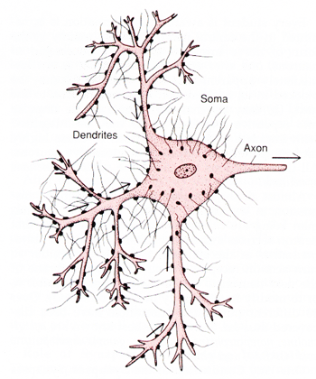
A typical motor neuron has 3 major parts
- Soma: main body
- Axon: extends from soma into a peripheral nerve leaving the spinal cord
- Dendrites: branching projections of the soma extending into surrounding areas of the cord.
Neurons vary in size and diameter. Some have an extra myelin sheathing around the axon, thought to enahance transmission speed.

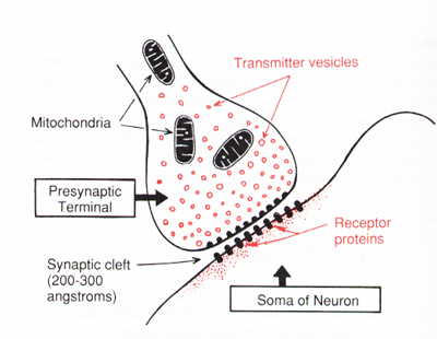
Presynaptic Terminals (aka terminal knobs, synaptic knobs): small knobs on dendrite and soma surfaces, which are the ends of nerve fribrils originating on other neurons. Contain transmitter vessicles which in turn contain transmitter substance which are released into the synaptic cleft between presynaptic and postsynaptic neurons. Substance may be excitatory or inhibitory to the postsynaptic neuron. Also contains mitochondria, which supply ATP (energy) to synthesize new transmitter substance.

The Process
AP depolarization over presynaptic terminal
-> vesicles empty into cleft
-> tranmsitter binds with receptor on postsynaptic neuron
-> postsynaptic neuron responds accordingly
Presynaptic Terminals: membrane contains voltage-gated calcium channels, which open during depolarization allowing calcium ions flow into the terminal. It is believed Ca2+ binds with proteins on the inner surface of the presynaptic membrane (release sites), causing local transmitter vesicles to bind and fuse with the membrane and open to the exterior via exocytosis. The quantity of transmitter released into the synaptic cleft is directly related to the number of calcium ions entering the terminal.
Receptor proteins in the postsynaptic neuron are composed of a binding component protruding into the synaptic cleft and an ionophore component that passes through the membrane to the interior. Two types of ionophores:
- Ion Channel: allows specific ions to pass.
Channel opening and closing is a rapid response to the presence
and then absence of transmitter substance.
- Cation channels: most often sodium, sometimes potassium or calcium. Opening of sodium channels excites the postsynaptic neuron, so transmitter substances opening these channels are excitatory.
- Anion channels: mainly chloride, but also minute quantities of other anions. Opening of chloride channels inhibits, and transmitter substances opening these are inhibitory transmitters.
- Second Messenger: activator protruding into the cytoplasm to activate one or
more substances in the neuron. Promotes prolonged changes in the neuron.
Typically a group of proteins attached to a receptor are activated. A portion
of this group separates and moves into the cytoplasm, where it can:
- open specific ion channels for a prolonged period.
- activate metabolic machinery that can in turn initiate other chemical results, including long-term changes in the cell structure.
- activate one or more intracellular enzymes, initiating specific chemical functions.
- activate gene transcription, forming new proteins in the neuron, which can change the metabolic machinery or structure of the cell.
Synaptic Transmitters
More than 50 identified transmitters are classified into two groups:
- Small Molecule, Rapidly Acting:
most acute responses such as sensory signals to the brain and motor signals
back to the muscles.
- Acetylcholine: secreted in many areas of the brain. Mainly excitatory, but inhibitory at some peripheral parasympathetic endings, such as heart inhibition by the vagus nerves.
- Norepinephrine: secreted by neurons with cell bodies in the brain stem and hypothalamus. Mostly excitatory.
- Serotonin: inhibitor of pain pathways in the cord. Also believed to help control mood and cause sleep.
- Dopamine, Glycine, Gamma-aminobutyric acid (GABA) all considered inhibitory, Glutamate mostly excitatory
- Neuropeptides:
cause more prolonged actions such as long-term changes in the numbers of
receptors, long-term opening or closure of certain channels, and possibly
the numbers and sizes of synapses. Synthesized along with new vesicles in
the soma. Examples include...
- Hypothalamic Releasing Hormones: cause pituitary to release its hormones into the circulation.
- Pituitary Peptides: released into posterior pituitary
- Sleep Peptides: released into basal regions on the brain to promote sleep.
Discharges by the postsynaptic neuron become progressively less when subjected to repetitive stimulation. This fatigue of synaptic transmission is typically caused by exhaustion of stores of transmitter substance in the synaptic terminals. Transmitter can be exhausted in only a few seconds to a few minutes of rapid stimulation. A protective mechanism -- when areas of the nervous system become overexcited, fatigue usually causes them to lose this excess excitability after awhile.
|
Intro |
Synapses |
Receptors & Circuits |
|
Motor Function |
Autonomic System
|