GE 345: Week 4
Myofibrils & NM Interaction
| Potential | Skeletal Muscle | Myofibrils, NM Interaction | Contraction | Fun Facts | |
Neuromuscular Interaction
Large, myelinated nerve fibers originating in the anterior horns of the spinal cord innervate skeletal muscle. Each nerve fiber branches to stimulate a few or hundreds of muscle fibers. Nerve endings form the neuromuscular junction at the muscle fiber's midpoint, with action potentials travelling outward toward the muscle fiber's ends. By and large, there is only one neuromuscular junction per muscle fiber. One motor nerve and all of the muscle fibers it innervates is known as a Motor Unit.
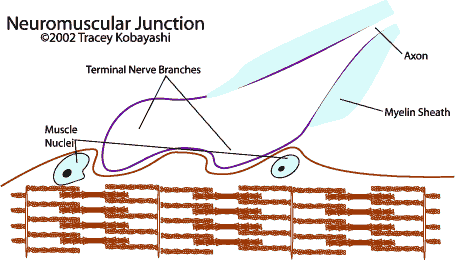
Acetylcholine (ACh), an excitatory neurotransmitter, is synthesized in the neuron. When a nerve impulse reaches the NM jct, ACh is released into the synaptic space. ACh opens a channel for ions to diffuse through at the motor end plate, beginning the AP in the muscle fiber. The ACh is destroyed by the enzyme acetylcholinesterase after a short time to end ion diffusion at the motor end plate.
The Myofibrils
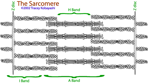
Actin filaments are attached to a Z-disc, a latticelike structure that forms the foundation for the molecular mechanism of shortening in muscle. The functional unit of the muscle is the sarcomere, which is the area from one Z-disc to the next. Actin and myosin give muscle its striated appearance when viewed under a light microscope or magnifying glass:
- I (isotropic) band: appears lighter because thin actin filaments allow more light through. The I band can disappear when the fiber is completely contracted, as the actin overlaps the myosin.
- A (anisotropic) band: darker because thicker myosin blocks more light.
- H band in the center of the A band appears where the myosin filament is less dense at its center.
Myosin is composed of myosin molecules, which are polypeptide chains. One end of the chain forms a head with a hinged arm, allowing it to extend from or move in close to the myosin filament's body. The head cleaves ATP, which releases energy for contraction.
Actin is composed of actin, tropomyosin and troponin. ADP molecules attached to certain actin molecules are believed to be the sites with which the myosin filaments interact.
| Potential | Skeletal Muscle | Myofibrils, NM Interaction | Contraction | Fun Facts | |