GE 345: Week 4
Muscle Contraction
Updated by Tracey 2 August 02
| Potential | Skeletal Muscle | Myofibrils, NM Interaction | Contraction | Fun Facts | |
The Contraction Process
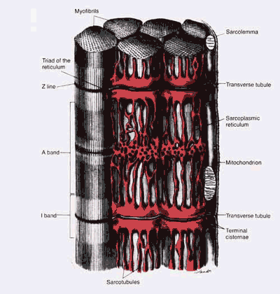
Action potentials travel along sarcolemma through T-tubules. Terminal cisternae of the SR adjacent to the T-tubule releases Ca2+, which penetrates the myofibrils.
Without the presence of troponin & tropomyosin, actin and myosin molecules bind instantly and strongly. When Ca2+ penetrates the myofibrils, it is believed to bind with the troponin on the actin chain. The troponin-Ca2+ complex cause the tropomyosin to shift, allowing interaction of actin & myosin. The myosin heads "ratchet" along progressive binding site of the actin. It's believed the cleaving and reformation of ATP provide the "power stroke" for this ratchet effect.
The Ca2+ is pumped back into the lateral sacs of the SR to promote relaxtion. When the troponin-Ca2+ complex is broken up, the tropomyosin shifts back to cover the binding sites.

Smooth Muscle Contraction
Smooth muscle in organ and vascular walls helps maintain their integrity, and functions to move contents along. Actin and myosin in smooth muscle interact similarly to skeletal muscle, and are activated by calcium ions as is skeletal muscle, however, there are some differences.
Smooth muscle doesn't have the same striated arrangement as skeletal muscle. Actin is attached to bodies that may be attached to the cell membrane, and myosin is interspersed among the actin filaments.
Most smooth muscle contraction is prolonged, lasting hours or days. Cross-bridge cycling is much slower, and attachment time of actin and myosin greatly increased. Much less energy is required to maintain smooth muscle tension.
| Potential | Skeletal Muscle | Myofibrils, NM Interaction | Contraction | Fun Facts | |